About Root Fracture SOS Clinic
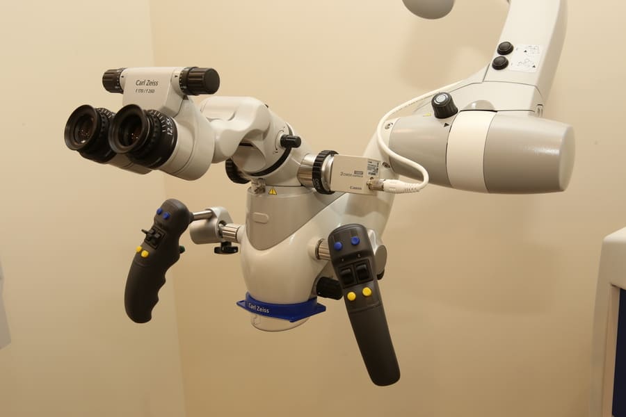
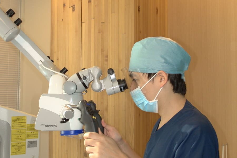
In our clinic, we conduct challenging treatments such as repair of “root fractures (cracks in the tooth roots)” and “perforations (holes) within the root canal,” as well as the removal of “fractured instruments within the root canal” using a dental operating microscope.
What is a tooth root?
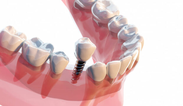
The term “tooth root” literally refers to the roots of the teeth. Analogous to the foundation of a house, it contains narrow tubes called “root canals,” where nerves and blood vessels are housed. These root canals have complex shapes, often curving or branching like the limbs of a tree. Bacterial infections from cavities can spread to these root canals, leading to the accumulation of pus at the tip of the tooth root. This can cause intense pain and swelling even in teeth without nerves.
Tooth root trouble
“Root fracture” refers to a condition where cracks or fractures occur in the tooth roots.
Traditional root canal treatments often struggled to thoroughly remove infectious materials from complex-shaped root canals, leading to the risk of leaving behind sources of infection. Even after initially cleaning and restoring with dental restorations, the root tip could become infected later, resulting in the eventual loss of the tooth, necessitating further root canal treatments.
To avoid such retreatments and preserve teeth as much as possible, our clinic recommends precise endodontic treatment using an operating microscope, in addition to extensive experience and proven techniques.
The use of an operating microscope in endodontic treatment began to spread in the 1990s, primarily in Western countries.
By providing a bright and enlarged view, it transformed endodontic procedures from relying on “intuition” and “manual dexterity” to precise and reliable practices.
About Root Fractures (Cracks in the Tooth Roots)

The leading causes of tooth loss are
- periodontal disease (37.1%)
- caries (29.2%)
- fractures (17.8%)
- other reasons (7.6%)
- impacted teeth (5.0%)
- orthodontics (1.9%)
- cause unknown (1.4%)
Considering that “fractures” rank third, many people may have experienced fractures. However, there is currently no established treatment for root fractures, leading to the typical diagnosis of “extraction.” While this is not an incorrect judgment, patients may be wondering, “Is extraction really the only option?” While it may not be possible to save all fractured teeth, our clinic employs a reinforcing method using special adhesives and glass fibers to pursue conservative treatment.
About Perforations (Holes) within the Root Canal
What is “perforations” ?
Perforations” occur when a hole forms in the root canal, allowing communication between the oral cavity and the surrounding tissues due to various reasons.
Many cases arise as accidental accidents during root canal treatment, but there are also cases caused by pathological factors such as the progression of caries or root resorption.
Difficulties in perforation treatment
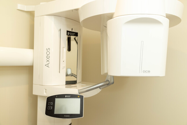
Confirming perforations within narrow root canals, especially in curved canals or near the root tip, can be challenging.Therefore, for confirmation, we use an operating microscope, magnifying glasses, and dental X-ray CT examinations.The location and size of perforations vary, and similar to “tooth fractures,” many cases are diagnosed as requiring extraction.
However, our clinic is committed to conservative treatment by sealing perforations.
“Before Starting Endodontic Treatment”
Before initiating endodontic treatment, crowns (restorations), fillings, and foundations attached to the teeth are generally removed.
In cases where root fractures or extensive cavities are identified during treatment or if there is a perforation in the tooth structure, the treatment plan may be changed to surgical endodontic therapy.
In cases where treatment is deemed difficult, extraction may become necessary.
Please note that the repair of “root fractures” and “perforations within the root canal” is considered a private treatment.
However, the removal of “fractured instruments within the root canal” may be covered by insurance in some cases.
Case
1. Upper Second Premolar Root Fracture
Root fractures (cracks in the tooth roots) are common occurrences. Our clinic reinforces root fractures using a method similar to the seismic reinforcement sheet method for tunnels. We use MMA-based dental adhesive resin cement (Super Bond C&B®, Sun Medical, resin cement) and glass fibers to reinforce the cracks. The procedure involves removing as much infectious material as possible within the fracture line, pouring resin cement after enlargement, and firmly adhering a cut glass fiber sleeve to the shape of the root canal.
Figure 1.
Vertical fracture of tooth root
Figure 2.
Repairing the fracture with a glass fiber sleeve
2. Removal of broken instruments inside the root canal
For the removal of foreign objects (intracanal foreign bodies) remaining within the root canal, dental X-ray CT imaging is first performed. Based on the image diagnostic results, we use an operating microscope for precise removal.
【Notice】
“Removing intracanal foreign bodies may result in a reduction in the thickness of the tooth, increasing the risk of accidental events such as perforation (the formation of a hole).”
Preparation for endodontic treatment
1. Gingival Cord Packing:
Gingival cord packing is a technique involving the insertion of a thin thread (packing cord) into the gingival groove (a physiological groove between the tooth and the gums).
This reveals the boundary between the tooth and the gums, eliminating the thickness of the gums equal to the thickness of the thread.
Without gingival cord packing, an accurate “isolation barrier” in the next stage cannot be achieved.
2. Isolation Barrier:
An “isolation barrier” refers to the creation of a wall on the tooth during treatment. Without creating this barrier, the next stage of “rubber dam” cannot be performed. Teeth without an isolation barrier may have a higher chance of not being able to start the root canal treatment, especially in the case of posterior teeth. In our clinic’s root canal treatment, we always create an isolation barrier, and for temporary sealing, we use light-curing composite resin (synthetic resin of the same color as the tooth). “Actual partition wall creation”
Figure 1.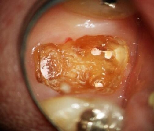 No remaining tooth structure on the gingival edge
Figure 2. Creating and bonding the isolation barrier
3. Rubber Dam:
The “rubber dam” is a rubber sheet that is essential for root canal treatment. It is highly effective in preventing saliva containing bacteria from flowing into the root canal, preventing reinfection.
It also ensures safe treatment by preventing accidental ingestion or aspiration of the drugs used during treatment.
Please tell us if you have a latex allergy (as anaphylactic symptoms may occur, and we will switch to a non-latex sheet).


